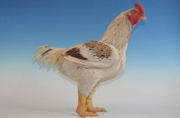
Gallus gallus
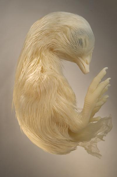
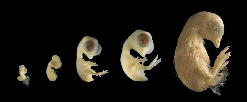
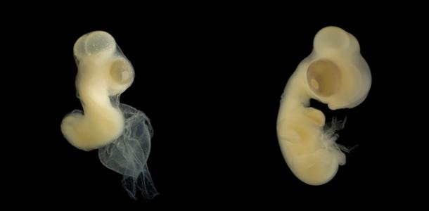
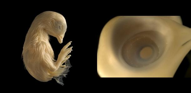
The Chicken embryo is a staple educational tool in developmental biology. Their availability and similarities with mammalian embryo, help shape our present understanding of embryology. After 21 days of incubation, the chick attempts to break out of its shell, pushing its beak through the air cell. Since the specimens were received out of the egg and without its yoke, I lacked the ability to document the chicken’s interaction in its element. The specimens document a range from 5, 6, 9, 12, to 18 days of development.
PHOTOGRAPHING THE EMBRYO
SUPPLIES
Camera
Two sets of fiber optic lights
Glass cell approx. 5″x5″
Water that has sat out in the open for 1 day
Black velvet fabric large enough to cover the bottom of the glass cell.
PROCEDURES
The chicken embryo is a semi-transparent and monotone subject. The transparent flesh lends itself well to back light, which causes the skin to appear to glow. The choice of background is a crucial prop that helps determine the success of the image for its application. For scientific and documentation purposes, black is the best choice to contrast the flesh tone embryo and emphasize its subtle details. Using a white background makes the translucent skin difficult to distinguish between the subject and its background. However, done correctly, a white background can produce an aesthetically pleasing photo, articulating its neutral tones.
The chicken embryo if filled with fluids. If taken out of the water, its delicate cavities will collapse, loosing the significance of its bodily form. Photographing the embryo through water maintains its structure. A second advantage of photographing the embryo under water removes any specular highlights while diffusing the light that falls on the subject. However, the diffusion does reduce the contrast. Bumping up the contrast either through the lighting technique or through the digital file will be helpful.
In the same way that air bubbles form on the sides of an open bottle of water, bubbles will form on the embryonic body. This produces unpleasant artifacts on your image. To reduce this issue, let the water sit out for a day so the gas in the water have a chance to escape. If the bubbles still remain on your subject, try shaking it off, otherwise as a final attempt, they can be removed through Photoshop. Fill the cell with water so it just covers the embryo. Too much water and the embryo will float around uncontrollably. Too little water and unwanted specular highlights will show up on its wet surface.
Place the glass cell on top of the black velvet. Black velvet produces a rich black background. Set the fiber optic lights at the bottom of the cell on two opposite sides of the cell. My lighting ratio was at 1:2. One light shining towards the front of the embryo and the other shining on the back. The head of the chicken is usually the main focus point. Let the brighter light shine on the anterior body. Placing the lights at the very bottom of the cell allows the light to shine across and through the transparent embryo, creating a glow on its edges. The side lighting also forms shadows on the embryo, emphasizing its bulbous eyes and porous skin.
Chicken embryos are a fascinating subject both on a scientific level and aesthetical level. The continual observation and documentation of chick embryo helps to educate the development of organisms generate and are manipulated.
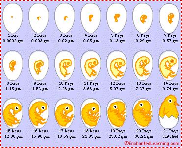
| . ………………………………. | DAY 5 Reproductive organs begin to form The bones of the legs & the crop begin to develop. |
……………………………… | DAY 6 Wing is bent at the elbow The beak is more prominent No egg tooth is visible yet |

| ………………………….. | 10 DAY The distal segments of limbs are longer Nostril is a narrow slit Flight feathers are noticeable Lower eyelid has grown up to level of cornea Circumference of lids is a narrowing ellipse |
……… | 12 DAY Primary scales form over entire surface of leg Feather germs surround auditory opening Upper eyelid is covered with feather germs Lower eyelid covers 2/3 to 3/4 of cornea |
18 DAY – Beak length-4.8mm – Third toe length-16.7mm
Facts sheet reference: http://www.rit.edu/~gtfsbi/genbiol/chicklab.htm
PHOTOGRAPHY & COPY PRODUCED BY MICHELLE LEUNG –
contact – michelleleung8@gmail.com
Egg and Embryo Development
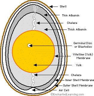
Cross Section of a Newly Laid Egg
The Formation of an Egg:
The Yolk: The chicken egg starts as an egg yolk inside a hen. A yolk (called an oocyte at this point) is produced by the hen’s ovary in a process called ovulation.
Fertilization: The yolk is released into the oviduct (a long, spiraling tube in the hen’s reproductive system), where it can be fertilized internally (inside the hen) by a sperm.
The Egg White (albumin): The yolk continues down the oviduct (whether or not it is fertilized) and is covered with a membrane (called the vitelline membrane), structural fibers, and layers of albumin (the egg white). This part of the oviduct is called the magnus.
The Chalazae: As the egg goes down through the oviduct, it is continually rotating within the spiraling tube. This movement twists the structural fibers (called the chalazae), which form rope-like strands that anchor the yolk in the thick egg white. There are two chalazae anchoring each yolk, on opposite ends of the egg.
The Eggshell: The eggshell is deposited around the egg in the lower part of the oviduct of the hen, just before it is laid. The shell is made of calcite, a crystalline form of calcium carbonate.
This entire trip through the oviduct takes about one day.
Growth of the Embryo: The fertilized blastodisc (now called the blastoderm) grows and becomes the embryo. As the embryo grows, its primary food source is the yolk. Waste products (like urea) collect in a sack called the allantois. The exchange of oxygen and carbon dioxide gas occurs through the eggshell; the chorion lines the inside surface of the egg and is connected to the blood vessels of the embryo.
The Incubation Period: The embryo develops inside the egg for 21 days (the incubation period), until a chick pecks its way out of its eggshell and is hatched.
Definitions:
Air Cell – an empty space located at the large end of the egg, it is between the inner and outer shell membranes.
Chalaza – a spiral, rope-like strand that anchors the yolk in the thick egg white. There are two chalazae anchoring each yolk, one on the top and one on the bottom. (The plural of chalaza is chalazae.)
Germinal Disc or Blastodisc – a small, circular, white spot (2-3 mm across) on the surface of the yolk, it is where the sperm enters the egg. The nucleus of the egg is in the blastodisc.
Inner Shell Membrane – the thin membrane located between the outer shell membrane and the albumin.
Outer Shell Membrane – the thin membrane located just inside the shell.
Shell – the hard, protective coating of the egg. It is semi-permeable; it lets gas exchange occur, but keeps other substances from entering the egg. The shell is made of calcium carbonate.
Thick Albumin – the stringy part of the egg white (albumin) located nearest the yolk.
Thin Albumin – the watery part of the egg white (albumin) located farthest from the yolk.
Vitelline (yolk) Membrane – the membrane that surrounds the yolk.
Yolk – the yellow, inner part of the egg where the embryo will form. The yolk contains the food that will nourish the embryo as it grows.
http://www.microscopy-uk.org.uk/mag/artnov04macro/mlchicken.html